This post continues with our occasional series looking at the normal functioning of the body. This follows the post a few days ago where we looked at the normal structure and function of the heart and how blood flows through the chambers of the heart and around the body and lungs. Here we consider the electrical nature of the heart muscle, how the ECG (EKG is the same thing) is generated and what the different wave forms mean in physiological terms.
A normal ECG (EKG) recorded from one of my students.
The components of the cardiac cycle and the events the electrical activity represents.
The internal electrical conducting system
The cardiac cycle is controlled by specialised conducting tissue in the heart. Inside the right atrium is an area of specialised cardiac muscle tissue termed the sinoatrial (SA) node. Because this controls the pace of the heart it is sometimes called the pacemaker. This area generates the initial electrical impulse which stimulates myocardial contraction. From the SA node an impulse spreads to both atria stimulating their contraction. The impulse travels across the atria via specialised conduction pathways termed the internodal tracts; this is because they are between the SA node and the atrioventricular (AV) node.
The AV node collects an impulse from the atria and passes it on to the bundle of His (or atrioventricular bundle) in the cardiac septum. The AV node is the only pathway the impulses can travel in order to spread from the atria to the ventricles; the rest of the tissue in the plane of the valves is electrically insulating. In the cardiac septum the bundle of His divides into two, forming the right and left bundle branches, which carry the electrical impulse to the right and left ventricles. The result of this arrangement is that an impulse is carried rapidly down through the cardiac septum. Finally the impulse innervates the ventricular myocardial muscle via the small Purkinje fibres (or conduction myofibres). This means that ventricular contraction will start from the cardiac apex and work towards the base, pushing the blood upwards, towards the arterial valves. It is this internal conducting system which is responsible for the initiation and phases of the cardiac cycle.
Unlike other muscles the myocardium internally generates the electrical impulses which lead to muscle contraction. As mentioned, this electrical activity originates from the sinoatrial node. However, outside factors will influence the heart rate and strength of contraction. Adrenaline will increase heart rate, as will stimulation by the sympathetic nervous system. This is why heart rate, and the strength of contractions, will increase during exercise, excitement or as a result of anxiety. Parasympathetic stimulation will slow the heart rate and so reduce cardiac output. When we are relaxed the parasympathetic nervous system will slow the heart rate and reduce the strength of individual contractions. The internal generation of the electrical activity required for cardiac contraction explains why a donated heart will carry on contracting after a heart transplant operation.
The components of the cardiac internal conducting system. The sinoatrial node generates a new electrical impulse prior to every cardiac cycle. Firstly, this impulse causes atrial contraction. As an electrical impulse cannot be transmitted through the non-conducting fibrous tissue of the valvular plane it passes down to the ventricles via the AV node. This same impulse then causes ventricular contraction.
The PQRST as seen using an electrocardiograph
When the myocardium is stimulated by an electrical impulse the myocardial cells will depolarise. This means the electrical polarity across all of the individual cell membranes will reverse. At rest any muscle cell is negatively charged on the inside and positive on the outside. Arrival of an electrical impulse will reverse this resting potential causing the cells to depolarize, becoming positive on the inside and negative on the outside. It is this depolarisation of the myocardial cells which initiates their contraction. The collective electrical activity of the myocardial cells depolarizing, and then repolarizing, may be detected with electrodes on the surface of the body. In health the contraction of the myocardial muscle cells will occur essentially at the same time as the cells depolarize. This means we can directly relate the electrical patterns we can detect on the surface of the body with the contractions of the myocardium. This is the principle of the electrocardiogram (ECG). When this is recorded three characteristic electrical phases can be clearly seen.
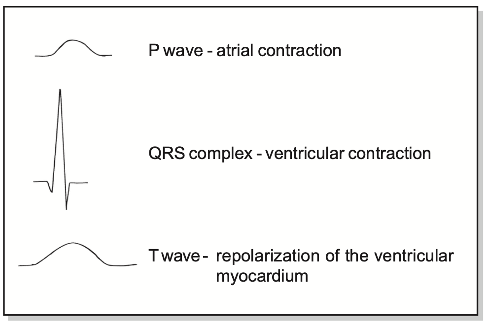
Firstly there is a P wave. This is the electrical activity, as detected on the surface of the body, as a result of the depolarization of the atrial myocardium.
Secondly there is the larger QRS complex caused by the depolarization of the larger muscle mass of the ventricular myocardium. This complex is associated with ventricular contraction. Thirdly there is a T wave. This is not associated with any muscular contraction but arises as the ventricular muscle repolarizes to an electrically resting state. Finally, there is a short gap before the next atrial contraction at the start of the next cardiac cycle.
A normal cardiac cycle must have a PQRST phase in that order. In health the occurrence of these phases of the cycle is fairly regular and the rate is usually between 60 and 100 per minute. So a normal rhythm has a PQRST, in the correct order, is regular, with a rate between 60 to 100 cycles per minute. This normal rhythm is called a sinus rhythm because the cardiac rhythm is controlled by the sinoatrial node in the right atrium.
If the phases of the cardiac cycle occur regularly, and in the correct order, at a rate of less than 60 times per minute, the rhythm is termed sinus bradycardia. This is normal in people who are physically fit or who are very relaxed. If the rate is over 100, with a regular PQRST in the right order, this is termed a sinus tachycardia. A sinus tachycardia is of course normal during exercise. The terms P,Q,R,S and T do not stand for anything and have no intrinsic significance what so ever. They are arbitrary names given to specified phases.
Effects of exercise
Exercise increases heart rate, which is the number of times the heart beats per minute. It also increases stroke volume which is the amount of blood pumped out per cardiac contraction. These two factors combine to increase cardiac output, which is defined as the volume of blood pumped out from the left ventricle per minute. To be precise, cardiac output equals heart rate multiplied by stroke volume. At rest a normal stroke volume will be around 70mls. If the heart rate is 72 beats per minute this would give a cardiac output of 70 x 72 which equals a cardiac output of 5040 mls. As an average adult has about 5 litres of blood in total, the cardiac output figure means the entire volume of the blood circulates through both the body and lungs once per minute.
With increasing levels of physical activity cardiac output will progressively rise. This will increase the rate at which blood circulates around the lungs and body tissues and so increase the delivery of oxygen and nutrients to active muscles. During vigorous exercise an average adult might be able to increase their cardiac output to 20 or 25 litres per minute for a period of time. A trained athlete will be able to achieve a cardiac output of 35 or even 40 litres per minute for a short time.
Regular exercise is good
Regular exercise is very good for humans; it lowers the levels of sugar (glucose) in the blood and increases levels of the protective HDL (high density lipoprotein) cholesterol. Exercise will increase metabolic rate and sustained exercise will burn up excess body fat preventing obesity. Although exercise raises blood pressure at the time, it lowers blood pressure overall. It makes the heart muscle stronger and helps to keep the coronary arteries patent. These factors mean regular exercise helps to protect against heart attacks and strokes. Recent findings indicate that regular exercise reduces the risks of developing some forms of cancer. Exercise tones and strengthens many muscles in the body and as exercise applies forces through the bones of the skeleton it will increase bone strength. Exercise in childhood and young adult life will build up bone mass and make osteoporosis less likely in later life. Regular exercise is an effective treatment for depression. If people are immobilised and unable to exercise, they may suffer from numerous complications such as blood clots in the veins of the legs and lungs, pressure sores, depression, constipation, pneumonia and atrophy of bones and muscles. This is why everyone should try to exercise for a least half an hour every day unless there is some medical reason not to.


Hello John, I've been following you since you started posting on Covid back in early 2020. Your info is top notch, but honestly it's your truly comforting wit and charm that keeps me coming back for more. If it wasn't for you and Tucker Carlson, I think i would have gone crazy! So thank you.
I was wondering if you'd considered doing a video about Hypochlorous Acid (HOCl) for use disinfection, antisepsis, and wound care? I believe they have found a way to stabilize it and extend the shelf life so now it is available everywhere, including Amazon in sizes up to a gallon (I discovered this morning, to my surprise). When i searched 'duck duck go' for Pure Hypochlorous Acid I came across a WHO link of a PDF titled 'Application for Inclusion in the 2021 WHO Essential Medicines List'. If you have a spare moment, please take a look at the submission. It's truly amazing! Kills just about everything on surfaces and topically with no resistance build up and no side effects and no PPE required. This could help a lot of people because it's cheap and effective and I hope you read this message and take a look. Dr. John Campbell, you are always in my prayers and thank you for putting up with so much bs to keep us all informed with our spirits lifted! Love ya!
Very interesting, thanks. Obviously you've been a great and respected teacher.