


The importance of cells
Structure and a few functions

One of the good things about using Substack is it gives you plenty of space to put in plenty of words and pictures. You as the reader of course can skip any individual posting or choose to spend a bit of time. This posting is for those who would like to know a little more about the biology of cells, how they fit into the hierarchy of the body, and why they are important to understand as part of the essential underpinnings of all biology.
Introduction
Before we get down to some really interesting physiology it may be useful to define a few terms. Physiology is the study of the normal function of a biological system, in this case the human body. Anatomy is the study of normal structure. Inevitably, in order to understand physiology, we must also learn some anatomy. Histology is the study of tissues and cytology the study of cells. Biochemistry is the study of the chemistry of living things; most of this chemistry takes place within individual cells. Pathology is the study of abnormal anatomy and pathophysiology means the study of abnormal body function. Histopathology studies abnormalities found in tissues. Psychology studies the normal processes of the mind. Psychiatry is the study of the abnormal mind.
Cells, tissues and organisation
The body is a remarkably complex structure which performs thousands of physiological functions. This means that the structures of the body must be organised in a precise way to carry out these functions. The functional systems of the body, such as the nervous or digestive systems, are themselves made up of organs such as the brain or stomach. Organs in turn are composed of precise arrangements of groups of tissues. A tissue is a group of similar cells, often with associated additional structural material produced and secreted by the tissue cells. Cells which compose a particular tissue are themselves composed of smaller functional units called cell organelles. These organelles can be thought of as the ‘organs’ of a cell as they perform specialised intracellular functions. Cell organelles are made up of highly structured biomolecules such as proteins, carbohydrates and fats. Large biological molecules are made up of smaller organic compounds. (Organic simply means that the molecule contains some carbon.) The organic molecules are composed of structured arrangements of atoms, which are composed of protons, neutrons and electrons.

Table 1.1
The body is very highly organised on a number of levels. This table indicates the hierarchy of this organisation, starting with the largest and working down to the smallest.
Cell organelles
Cells may be considered as the ‘units of life’. This means that many of the processes essential for life are carried out inside cells. Cytology is the term used to describe the study of cells. A single cell may be considered to be ‘alive’. Some simple organisms such as the amoeba are composed of a single cell. However, in humans billions of cells combine to make up the body. As cells are only 5-10 micrometres (a micrometre is one thousandth of a millimetre) in size they can only be seen with the aid of microscopes. Under a light microscope a cell has a dark staining central region called the nucleus with a clear area surrounding this, referred to as cytoplasm. These components are all surrounded by a cell membrane which encloses the cell.

Diagram 1.1
Diagram of a cell as viewed under a light microscope with a magnification of about 300 times.
However, when cells are examined under the higher power of an electron microscope the organelles may be seen. These are sub units or components of the cell. In the same way that a body is made up of organs a cell is composed of organelles. Organelles are structures within the cytoplasm which perform specific functions. They are the functional units of a cell.
Cell Membrane
This is a very thin membrane which surrounds the outside and marks the boundary of the cell. The cell membrane is composed of a combination of lipids, phosphates and proteins. Because the cell membrane is composed of two layers of phospholipid molecules it is referred to as a phospholipid bilayer. A phospholipid molecule is made up of a combination of phosphate and fatty acids. Cell membranes also contain some cholesterol. The external cell membrane compartmentalises the cell and controls movement of fluid and other substances in and out. This regulatory function means the cell is able to control, to a degree, its own internal environment.
Some of the proteins in the cell membrane are referred to as transmembrane proteins. This means they span the membrane and have ends in the external tissue fluid and internal cytosol. Some of these proteins regulate the passage of substances into and out of a cell. For example, some transmembrane proteins act as tunnels to allow glucose molecules from the tissue fluid into the cytosol. Other membrane bound proteins act as receptors. These specialised receptor protein molecules allow the cell to recognise chemical messages from other cells or from endocrine glands. Once a receptor has detected the presence of a particular chemical messenger this will initiate some physiological change within the cell. As a result cells do not function in isolation but as co-ordinated and disciplined components of the whole body.
Cytosol
Cytosol is the fluid found in the cytoplasm of the cell; it is the intracellular fluid. Cytosol is mostly water with some additional dissolved and suspended molecules, the combination of which is referred to as a colloid. Organelles are suspended within the cytosol and much of the water in the body is located in the cytosol. Many biochemical reactions occur in the cytosol such as the initial processing of glucose in the production of energy and the synthesis of fatty acids.
Cytoskeleton
Microfilaments and microtubules are composed of proteins and extend throughout the cytosol. Collectively these structures form an internal ‘skeleton’ within a cell referred to as the cytoskeleton. The cytoskeleton provides structural support and strength for a cell and helps to maintain overall shape. Cell movement is also facilitated by the cytoskeleton. This is necessary during the early embryonic development of the body where cells must migrate into the correct positions. Movement is also important for muscle contraction and the movement of white blood cells to combat infection.
Endoplasmic reticulum (ER)
This is an extensive network of flattened interconnected channels and tubules distributed throughout the cytoplasm. Endoplasmic reticulum is itself bounded by membranes. The function of this extensive structure is to move materials around inside the cell, i.e. intracellular translocation. As different organelles within a cell perform different functions, particular substances will be required in specific locations. In this sense the ER is analogous to a road or rail network, delivering raw materials and transporting away finished products. In addition the ER provides support for the overall structure and shape of the cell. The ribosomes are often associated with the endoplasmic reticulum. If an area of ER is associated with ribosomes it is described as rough ER, areas without associated ribosomes are termed smooth.
Ribosomes
These are small dark staining organelles. They are often associated with rough endoplasmic reticulum but free ribosomes may be found anywhere in the cytoplasm. Rough endoplasmic reticulum is described as ‘rough’ because it is studded with ribosomes. Ribosomes receive genetic instructions from the DNA (deoxyribonucleic acid) in the nucleus of the cell. This information is then transferred to molecules of RNA (ribonucleic acid) which then string together long chains of amino acids to form polypeptides and proteins. Amino acids are subunits of proteins in the same way that bricks are subunits of a wall. Ribosomes are therefore the site of protein synthesis, in essence they are little protein factories. Some of the synthesised proteins are used to form the structures of the individual cell and others are exported from the cell. It is logical that the ribosomes are usually associated with the ER so that they can be supplied with amino acids which are the building blocks of proteins. Synthesized proteins can then be removed to where they are required.
Golgi complex
This is an arrangement of membranes which are responsible for the export of products from a cell. Products from the ER are transported to the Golgi where some further molecular modifications often take place. Once the fat or protein based product is ready, so called secretory vesicles bud off the main Golgi complex and transport the material to the external cell membrane for export. For example, some cells in the digestive system secrete digestive enzymes in this way. Endocrine cells secrete endocrine hormones and liver cells secrete plasma proteins.
Mitochondria
All living cells must be able to produce energy to fuel physiological processes. When a cell is no longer able to produce energy it will quickly die. Mitochondria are usually sausage shaped and have an outer membrane and a highly enfolded inner membrane. This enfolded inner membrane provides a large surface area for the location of enzymes responsible for generation of energy within a cell. These enzymes produce energy by using oxygen to oxidise or ‘burn’ fuels such as glucose or fatty acids. The energy actually comes from breaking the chemical bonds which hold together the fuel molecule. All of the fuel required for energy production derives from food and all of the oxygen from breathing. Mitochondria are therefore the site of all energy production and are sometimes referred to as the ‘power house’ of the cell. Cells which use a lot of energy, such as those in muscles, have a lot of mitochondria in their cytoplasm whereas cells which need less energy, such as those which compose the skin, have fewer mitochondria. Cyanide is such a fast acting poison because it halts the process of energy production in mitochondria. Interestingly, mitochondria also contain some DNA. This mitochondrial DNA is only passed on through the maternal line, i.e. from mothers to daughters or from mothers to sons.
Lysosomes
Lysosomes are organelles surrounded by the typical membrane bilayer and contain a variety of digestive enzymes such as lysozyme. They are formed by breaking away from the Golgi apparatus and then dispersing throughout the cytosol. Lysosomes are used in the process of intracellular digestion, i.e. digestion within the cell. Digestive enzymes in the lysosomes break up large complex molecules into smaller ones using the process of hydrolysis. These hydrolysing type of enzyme split large molecules up by adding hydrogen and oxygen. This process of intracellular digestion may digest material from outside the cell. For example, in white blood cells foreign material is ingested by the cell, lysosomes approach the foreign material and spill their digestive enzymes onto it, therefore digesting it. Lysosomes are therefore essential for the process of phagocytosis, a term which literally means ‘cell eating’.
In addition to this digestion of extracellular material, lysosomes may also digest intracellular material, i.e. material derived from within the cell itself. As cells need constant maintenance, new components and organelles are constantly being formed meaning old ones need to be removed. Unwanted cellular organelles and biomolecules are digested by lysosomal enzymes in the process of autophagy. Lysosomes may also burst to digest a whole cell which is unwanted by the body in a form of cell suicide called apoptosis.
Peroxisomes
These organelles have a similar appearance to lysosomes but are smaller. Peroxisomes contain enzymes, including hydrogen peroxide, which work by oxidizing toxic substances. If these toxins were not oxidized they would build up to poisonous concentrations within cells. For example, about half the alcohol a person might drink is detoxified by peroxisomes in liver cells.
Nucleus
The nucleus is located at the centre of the cell inside the cytoplasm. There is a separate membrane surrounding the nucleus referred to as the nuclear membrane. Within the nucleus are the chromosomes. All human cells, (at least at some time in their lives) contain 46 of these bodies arranged in 23 pairs. Chromosomes contain the genes which are the genetic information responsible for making and controlling the cell. This is why the nucleus is sometimes referred to as the ‘control centre’ of the cell.
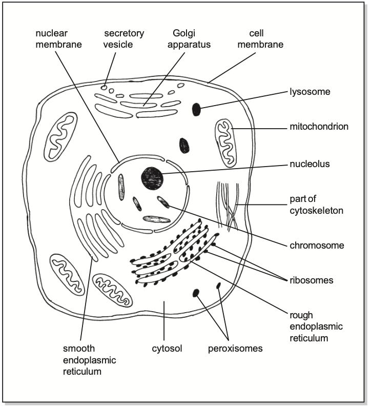
Diagram 1.2
Cell viewed under an electron microscope.
Chromosomes
The term chromosome literally means coloured body. The chromosomes are composed of structural proteins and DNA (Deoxyribonucleic Acid). Genes are part of the chromosomes and are composed of DNA strands. Genes contain the genetic information to create the cell and therefore ultimately to generate the body.
Nucleolus
A nucleolus is a cluster of DNA, RNA and protein within the nucleus. These are the same type of molecules found in the ribosomes. DNA carries the genetic code which is transcribed to form information carrying RNA molecules. This RNA is then used in the synthesis of protein which will form part of the ribosomes. Once formed these ribosomal proteins migrate out into the cytoplasm to form the ribosomes. During periods of increased protein synthesis, the nucleoli increase in size. They are also prominent in liver and muscle cells which synthesise large amounts of protein.
Specialised cells
During growth and development cells differentiate into specific types which allows them to perform particular functions. This differentiation is initiated and regulated by genetic instructions from the genes which carry all of the required information. Different cells must have a specialised structure which results in the production of many completely different types of cell. It is this process of cell specialisation which is referred to as differentiation. Specialised cells are required to form the different types of tissue needed to construct the large structures and organs of the body. Different types of cells include nerve cells or neurones, muscle cells, liver cells, blood cells and epithelial cells. Each cell has a structure and function specific to the role that a cell or tissue is required to perform in the body. Groups of similar cells compose tissues. Groups of tissues compose organs and groups of organs compose the systems of the body.
Cell reproduction
All cells are derived from parent cells which underwent a process of cell division. In this process one parent cell grows then divides into two daughter cells. There are only two forms of cell division, referred to as mitosis and meiosis.
Mitosis
Mitosis is often described as ‘simple’ cell division. During the non-dividing phase of the cell cycle a new, duplicate, set of 46 chromosomes is produced. Each gene is precisely copied to make an exact copy of the original. During
mitosis one complete set of chromosomes migrates to each end of the cell. This parent cell then divides into two daughter cells each also containing 46 chromosomes. Because the daughter cells and the parent cell all contain the same number of chromosomes this form of cell division is sometimes referred to as conservation cell division (the number of chromosomes is conserved). The group of descendent cells from a parent cell is described as a cell line.
Mitosis is the type of cell division that occurs when the body grows from a zygote, which is a single fertilised cell, into a baby. It is also the form of cell division which generates new tissue in the process of wound healing. In addition ongoing mitosis maintains the health of most body tissues by replacing old cells with new ones. Because mitosis is the type of cell division that goes on in the body it is sometimes referred to as somatic cell division (soma means body).

Diagram 1.3
The stages of mitosis in order.

Diagram 1.4
The principle of conservation cell division. A parent cell with 46 chromosomes mitotically divides to produce two daughter cells also with 46 chromosomes. After formation the daughter cells will usually grow until they become the same size as the parent cell.
Meiosis
The other form of cell division is called meiosis and is only used in the production of gametes which are the male sperm and female ova. The function of sperm and ova is to fuse together, forming fertilised cells. Such a fertilised cell is called a zygote. Once formed, a zygote undergoes repeated mitosis to form a new body. If the sperm and ovum each contained 46 chromosomes then the zygote, and hence the new baby, would contain 92 chromosomes. To prevent this increase in chromosome number the sperm and ovum are produced by meiosis which is a reduction cell division. During meiosis the daughter cell (always a
sperm or ovum) only receives one from each pair of chromosomes, leaving each sperm or ovum with 23 chromosomes. This means the zygote receives 23 chromosomes from the ovum and 23 from the sperm to give it a full complement of 46. Gametes are produced from germ cells which are only located in the testes and ovaries.

Diagram 1.5
Meiosis; the number of chromosomes is reduced from 46 to 23 per cell.
Diagram 1.6
During fertilisation the sperm and the ovum combine to produce a zygote with 46 chromosomes.

Diagram 1.7
Human life cycle. A male gamete fertilises a female gamete to form a zygote. This divides by the process of mitosis eventually forming a full-term baby. After birth there is further mitosis resulting in the growth of the baby, through childhood, into adulthood. The adult then produces gametes, by the process of meiosis, to start the next generation.
Cells and ageing
It seems that many cells have a limited number of times they can divide. Many of these permissible cell divisions take place between the formation of the zygote and the birth of the individual. This means in childhood and adult life only a limited number of further cell divisions are possible. When this number has occurred the cells are not capable of further divisions which results in death of the cell line. If the number of viable cells in a tissue is reduced the result will be that the tissue as a whole deteriorates. As the body is composed of tissue, deterioration in tissues results in the changes in the body we associate with ageing. Ultimately tissues become so non-viable that they fail to perform essential physiological functions, possibly resulting in the death of the individual. For example, the tissues in the wall of a blood vessel may fail resulting in haemorrhage.
As cells age there may also be mistakes in the process of cell division. For example, mutation can occur as a result of a mistake in the production of a new gene or chromosome as a cell divides. The term mutation means a change in a gene or chromosome. This is analogous to copying out a page of writing and misspelling or missing out some words. The accumulation of these errors in the genetic material of the cell may be one reason why many cancers are more common in older people than in younger people.
Another possible reason why cells age is free radical theory. A free radical is a very reactive form of oxygen which can oxidise and so damage cellular proteins. If free radicals attack the DNA of the genes this may result in age related changes and sometimes mutations which can lead to cancer. While some free radicals are produced as a result of normal metabolic processes, increased numbers may be generated as a result of over eating, exposure to radiation or exposure to polluting chemicals. Some vitamins and other components in fruit and vegetables called flavonoids, inhibit the activity of free radicals and so may slow down cell ageing. This is one reason everyone is advised to eat at least five portions of fruit and vegetables per day.
As people age the rate of mitosis and tissue repair is also reduced as there is a generalised slowing in the rate of metabolic activity. This is one reason why wounds heal much faster in children than in the elderly.
I am sure that it is understood, like when employed by LGA one speaks LGA, when employed by BBC one speaks BBC, However, dearest Dr Campbell, you bring Dr speak down from the highest enabling it to be understood, importantly understood, by all whom may not be privileged or accustomed to such medical speak. As did Mr Einstein with his E=MC□ who knows such complicated equation otherwise!, So from Joe Ordinary...Many Thanx..🌟🫂🌟 just to assure you that you are very much appreciated in our world 🌎.
Thank you.
This is an important piece of information for many of those who are not trained in medicine or health related subjects.
Without understanding this basis, I can't explain to anyone why mitochondria are important, why they have their own DNA, that isn't inside the nucleus and is thus is susceptible to damage by foreign invaders and certain toxic proteins.
Nor the downstream consequences of these subtle changes in the energy producing organelles and how that effects cellular homeostasis and gene expression.
And without that understanding, people won't understand how the same mechanism can be responsible for so many horrible observations.
Like misfolded proteins appearing.
Or neuropathies, inflammation and cytokine production.
Or immune dysfunctions.
Cardiac inflammation.
And a host of other consequences.
Mitochondria are important little organelles.